optos retinal imaging

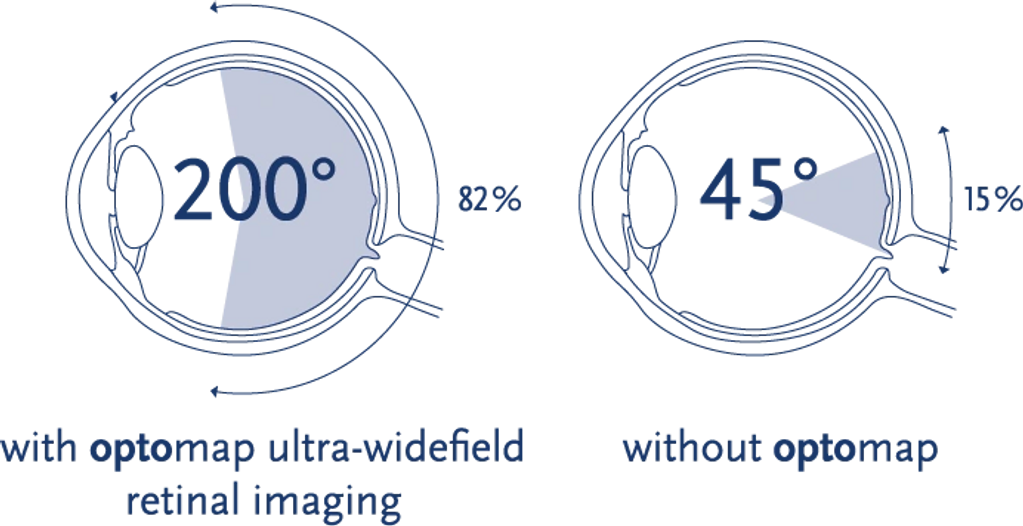
Mack Eye Center has the Optomap by Optos, which offers unparalleled views of the retina which provide eye care professionals with the following:
-The only anatomically correct 200⁰ or 82% image of the retina
-Simultaneous view of the central pole, mid-periphery and periphery

The Optomap clearly shows more Retinal anatomy than the traditional view, making for a faster and more thorough exam.

This is a normal retina as viewed through a non-dilated pupil.
optos and retinal disease

The dark cloud in the upper right corner is a large Retinal Detachment.

The yellow spots seens here are called Drusen, which are macular deposits that are seen with Dry Macular Degeneration.

Diabetic Retinopathy can be seen easily with the Optos; clusters of retinal bleeding are noted here.
Copyright © 2026 Mack Eye Center
(732)835-2020
This website uses cookies.
We use cookies to analyze website traffic and optimize your website experience. By accepting our use of cookies, your data will be aggregated with all other user data.